

Vertical root fractures (VRFs) are among the most difficult conditions for you to diagnose accurately in daily practice. Subtle signs and inconclusive imaging often delay confirmation until your patients’ prognosis is compromised. This blog is written for dentists who want to sharpen diagnostic accuracy when managing suspected VRFs.
You’ll learn why these fractures are often missed, how to interpret subtle clinical and radiographic patterns, the best treatment pathways, and how advanced dental continuing education at IDEA can enhance your fracture detection, case selection, and surgical or restorative planning skills to protect your patients’ long-term outcomes.
Diagnostic Challenges of Vertical Root Fractures
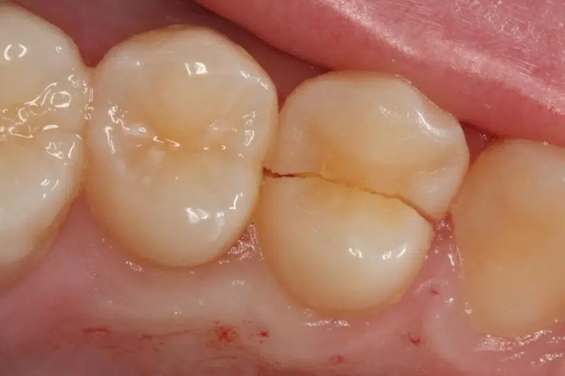
Vertical root fractures rarely announce themselves. Unlike acute pulpal pathology or classic periapical disease, VRFs tend to present through a collection of subtle, overlapping findings.
They can mimic endodontic failure, isolated periodontal defects, or combined lesions. For dentists managing these cases, the challenge is recognizing that a fracture may be present at all.
Several key challenges commonly arise when evaluating suspected vertical root fractures:
Symptoms That Point Everywhere and Nowhere
Patients with VRFs frequently describe mild biting discomfort, intermittent swelling, or dull, positional pain that does not fit a clean picture. These complaints overlap with occlusal trauma, periodontal abscess, and failing endodontic treatment. This variability can be misleading, making it crucial to consider VRF early to prevent unnecessary retreatment.
Dentists who can systematically evaluate the symptomatic patient are better positioned to catch VRF before misdiagnosis leads to the wrong treatment. IDEA’s Occlusion TMJ Bruxism Airway Dental Course helps you approach symptomatic patients with a functional perspective, improving your ability to distinguish fracture-related pain from other sources and supporting better long-term outcomes for your patients.
The Limits of Radiographic Evidence
Radiographs are a cornerstone of endodontic diagnostics, but they have inherent limitations, especially when it comes to detecting vertical root fractures (VRFs). Understanding these constraints helps dentists combine imaging with clinical examination for more accurate decision-making.
Key limitations of radiographic evidence:
- Beam alignment dependency: Fracture lines are only visible when the X-ray beam aligns with the fracture plane, which rarely occurs by chance.
- Indirect signs are inconclusive: Radiolucent halos, J-shaped lesions, or isolated crestal bone loss may suggest a fracture but are not definitive proof.
- Resolution limitations of CBCT: Cone-beam CT improves detection but hairline fractures can still be missed if they fall below the scanner’s resolution.
- Interference from metal artifacts: Existing restorations or posts can obscure fractures, making interpretation challenging.
- Need for clinical correlation: Radiographs alone cannot confirm VRFs; combining imaging with clinical signs, symptoms, and tactile assessment is essential for confident treatment planning.
Endodontic History Complicates the Picture

Teeth that have undergone endodontic treatment are the most common sites for vertical root fractures (VRFs). Symptoms often mimic standard endodontic failure, making diagnosis challenging. Recognizing unusual patterns can prevent unnecessary retreatment and preserve patient teeth.
Key insights for endodontically treated teeth:
- Misleading symptoms: Pain or infection in a treated tooth is often assumed to be persistent infection, missed canals, or inadequate obturation, while the underlying fracture goes unnoticed.
- Fractures often hidden: VRFs frequently remain undetected until surgical exploration or extraction reveals the true cause.
- Clinical red flags: Deviations from normal endodontic failure — such as unusual pain, mobility, or isolated bone loss — warrant further investigation.
- Value of advanced training: Dentists with specialized endodontic expertise can spot subtle signs and assess structural integrity more effectively.
- Hands-on skill development: The Endodontics Hands-On Course teaches dentists to evaluate complex cases, improving diagnosis, treatment planning, and long-term patient outcomes.
Narrow Pockets That Suggest Two Different Diagnoses
A deep, isolated periodontal pocket confined to a single root surface is one of the more reliable signs of VRF. It is also consistent with localized periodontal disease. Without surgical exploration, this distinction is difficult to make with confidence. Attention to subtle pocket morphology allows you to prioritize interventions that maintain periodontal health while addressing structural compromise.
Fractures That Propagate Before They Present
VRFs often begin as microscopic cracks that propagate slowly under occlusal loading. By the time symptoms are present, bone loss may already be established. Early-stage fractures leave no radiographic footprint, no probing anomaly, and no mobility.
The window for intervention narrows quietly. That is why a thorough functional and occlusal evaluation should be part of every diagnostic workup on a previously treated posterior tooth.
The Restorative Context as a Risk Indicator
Teeth restored with posts carry measurably higher fracture risk, particularly with rigid or oversized posts. When a post-restored tooth presents with nonspecific symptoms, post-related stress and fracture propagation should be evaluated in parallel. The presence of a post does not confirm a fracture, but it raises the index of suspicion enough to change the diagnostic approach.
No single finding confirms a vertical root fracture, and no single test rules one out. Accurate diagnosis depends on pattern recognition, a thorough case history, and the discipline to keep fracture on the differential even when other explanations seem more immediate.
Also Read: A Dentist’s Guide to the 5 Types of Tooth Cracks
Treatment Options for Vertical Root Fractures

Once a vertical root fracture is confirmed, treatment decisions depend on tooth type, fracture extent, periodontal involvement, and patient-specific factors. Early detection improves the likelihood of preserving structure and minimizing bone loss.
Extraction
For single-rooted teeth, complete vertical root fractures often necessitate extraction. Once the fracture extends through the root, bacterial infiltration along the fracture line compromises long-term prognosis. Extraction eliminates infection and prepares the site for future restorative planning, including implant placement or prosthetic solutions.
Root Resection or Hemisection
In multi-rooted teeth, such as mandibular molars, a fracture confined to one root may allow for hemisection or root amputation. By removing the affected root and preserving the remaining structure, you can maintain partial function. Case selection is critical, and periodontal stability must be assessed carefully.
Intentional Replantation with Bonding (Selected Cases)
In rare and carefully selected cases, intentional extraction, fracture repair, and replantation may be attempted. This approach remains controversial and technique-sensitive. Long-term prognosis varies significantly and requires precise case selection and advanced skill.
Guided Bone Regeneration After Extraction
When extraction is necessary, preserving alveolar bone becomes the next priority. Bone grafting and guided bone regeneration techniques help maintain ridge volume for future implant placement. Timely intervention reduces the risk of collapse and simplifies restorative planning.
Immediate Implant Placement
In certain scenarios where infection is controlled and bone conditions permit, immediate implant placement following extraction may reduce treatment time and preserve tissue contours. This option demands surgical precision and a comprehensive understanding of implant biomechanics.
Read more: Direct Composite Restorations in the Smile Frame
How Can You Help Prevent Vertical Root Fractures?
While not every vertical root fracture can be prevented, the way you perform endodontic and restorative procedures plays a major role in reducing the risk. Careful treatment planning, conservative techniques, and proper material selection can help protect the remaining root structure and improve long-term outcomes.
Use Conservative Post Placement Techniques
When placing a post after root canal therapy, preserve as much dentin as possible. Excessive canal enlargement during post preparation weakens the root walls and increases the likelihood of fracture over time.
Avoid Overusing Rigid Posts
Highly rigid posts can concentrate stress within the root. When appropriate, consider using fiber posts because their flexibility helps distribute occlusal forces more evenly along the root structure.
Preserve Tooth Structure During Root Canal Treatment
Removing excessive dentin during canal shaping can compromise the strength of the root. By maintaining adequate dentin thickness, you help the tooth better withstand normal chewing forces.
Adjust Occlusion When Necessary
After restoring an endodontically treated tooth, check the occlusion carefully. Reducing heavy contacts or excessive biting forces can lower the mechanical stress placed on the root.
Design Restorations With Adequate Ferrule
When placing a crown, aim for an adequate ferrule—generally around 1.5 to 2 mm of sound tooth structure above the crown margin. This helps distribute functional forces and strengthens the restored tooth.
Monitor Treated Teeth During Follow-Up Visits
Teeth that have undergone root canal therapy or post placement benefit from regular follow-up evaluations. Monitoring them during routine exams allows you to detect early signs of structural compromise before a fracture progresses.
Courses to Improve Diagnosis and Treatment Outcomes
Managing vertical root fractures effectively requires advanced training in endodontics, surgical techniques, restorative planning, and occlusion. IDEA offers hands-on dental CE courses designed to elevate confidence and improve patient outcomes.
Dental Microscope Course
Discover the power of magnification and precision in the Dental Microscope Course, which trains dentists to spot microfractures, crack lines, and subtle structural defects that can easily be missed. Improved visualization helps ensure more accurate diagnosis and treatment planning for challenging cases.
Bone Regeneration Hands-On Course
Develop hands-on expertise in regenerative dentistry through the Bone Regeneration Hands-On Course, focusing on bone grafting, membrane placement, and ridge preservation techniques. These skills are critical when preparing extraction sites of fractured teeth for future restorative solutions.
Immediate Implants Course
Learn strategies for efficient, predictable tooth replacement in the Immediate Implants Course, covering implant placement immediately after extraction, tissue management, and aesthetic planning. This course is ideal for dentists managing vertical root fractures where timely intervention is necessary to restore function and appearance.
Elevate Your Practice with Advanced Training

Vertical root fractures challenge even experienced dentists due to their subtle presentation and complex management pathways. Improving diagnostic accuracy and expanding surgical and restorative skillsets can significantly enhance patient outcomes while reducing retreatment and uncertainty.
Through world-class continuing education at IDEA, dental professionals gain practical, hands-on experience guided by internationally recognized experts. With limited seats available for many 2026 programs, now is the time to strengthen your diagnostic precision and surgical confidence.
Secure your place in an upcoming IDEA course and advance your ability to manage vertical root fractures with clarity, competence, and excellence.
